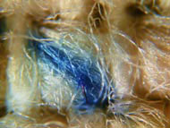
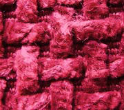
Contamination
The presence of contaminant materials in textiles
usually takes one of two forms; Either the unwanted component is
suspected in the raw material, which it will devalue, (e.g. wool
contamination in cashmere) or the contaminant passes unnoticed
through processing until the final product is dyed or finished,
where it becomes immediately obvious (e.g. polyolefin contamination
in natural fibre materials.
The presence of contaminants can devalue products
and materials to a significant degree. In textiles, such contaminants
may be incompatible fibres, raw material irregularities or foreign
matter. For other products, metals, plastic components, webs etc.,
the contaminant may be of an isolated particulate nature, or a
surface coating. Using light and electron microscopy such contaminants
may be identified and their likely origins suggested;
Examples of contaminants in textiles and other
materials may be;
Dark hairs, polyolefin packing materials,
vegetable matter, extraneous fibre types, non-fibrous matter, oxidation
products or moulds, extraneous coatings which impair processes
(e.g. cause dye resists), localised damaged areas and fugitive
tints.
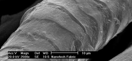
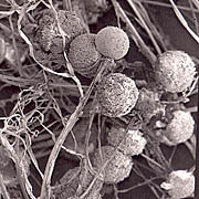
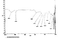
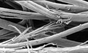
Micrography
For many reports or demonstrations, photo or
electron micrographs are and invaluable means of supplementing
written descriptions. A series of magnified images of a feature
or product can offer indisputable proof of the subject under study.
The understanding of an effect or a structure can be greatly enhanced
by having pictorial images to supplement written or spoken descriptions.
Light micrographs can show colour effects, internal structures
and reflected light features, while scanning electron micrographs
(SEM) are able to show surface features in great depth of field
and with superior resolution. The SEM can also be used to obtain
elemental information and it is possible to produce ‘x-ray
maps’ of the composition of a material or surface.
Areas where such pictures may be of value include;
Demonstration or teaching, legal evidence,
advertising and publicity, forensic science, report supplements. |